The EACR’s ‘Highlights in Cancer Research’ is a regular summary of the most interesting and impactful recent papers in cancer research, curated by the Board of the European Association for Cancer Research (EACR).
The list below appears in no particular order, and the summary information has been provided by the authors unless otherwise indicated.
Use the dropdown menu or ‘Previous’ and ‘Next’ buttons to navigate the list.
1. Mitotic clustering of pulverized chromosomes from micronuclei
Lin, Y-F. et al. Nature. 618: 1041-1048 (2023).
doi: doi.org/10.1038/s41586-023-05974-0.
Summary of the findings
Cancer genomes commonly harbor complex rearrangements driven by chromothripsis, the catastrophic shattering and reassembly of individual chromosomes. Chromothripsis can be initiated by mitotic errors that entrap mis-segregated chromosomes within micronuclei, which are susceptible to DNA damage and replication defects that lead to chromosome shattering in the subsequent mitosis. Most of the chromosomal fragments are then reassembled in random order to generate rearrangements. However, since most fragments lack a centromere and cannot attach to spindle microtubules, how shattered chromosomes undergo segregation during mitosis and reincorporation into daughter cell genomes was unknown. Lin et al. report that shattered chromosomes cluster in spatial proximity during mitosis, which biases fragment distribution toward one daughter cell. The authors identify the CIP2A–TOPBP1 complex as the mitotic tether, the inactivation of which disperses fragments throughout the mitotic cytoplasm.
Since shattered chromosomes are mostly inherited by one daughter cell, these findings predict that some cancer genomes would exhibit complex rearrangements lacking changes in DNA copy-number. Indeed, such copy-number-neutral rearrangements, termed balanced chromothripsis, are found in ~5% of diverse tumors and underpin the acquisition of recurrent driver events, including fusion oncogenes. This work highlights a mechanism that drives specific patterns of genome rearrangements from an initial chromosome segregation error.
CIP2A-TOPBP1-mediated mitotic clustering of pulverized chromosomes from micronuclei facilitates balanced rearrangements in one of the daughter cells following mother cell division. In the absence of CIP2A-TOPBP1, pulverized fragments disperse throughout the mitotic cytoplasm and stochastically partition into both daughter cells. The figure and figure legend were taken from Extended Data Figure 11 from the original article (https://www.nature.com/articles/s41586-023-05974-0#Sec30) published Open Access under a Creative Commons Attribution 4.0 International License (http://creativecommons.org/licenses/by/4.0/).
Future impact
It remains unclear what are the precise DNA lesion(s) that recruit CIP2A and TOPBP1 to micronuclei during interphase and/or to shattered chromosomes in mitosis. Additionally, further mechanistic studies are needed to determine how this protein complex functions to tether chromosome fragments, whether other unidentified components are involved, and how these processes are regulated in diverse contexts. Lastly, further investigation is needed to define the impact of balanced chromothripsis on cancer genome evolution and tumorigenesis.
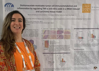
We caught up with Mounia Benbelkacem, a PhD researcher at the Laboratory of Cellular and Molecular Biology, Faculty of Biological Sciences, University of Science and Technology...
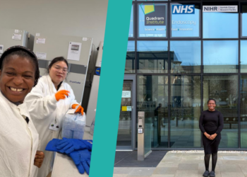
Aniefiok John Udoakang is a postdoctoral researcher at University of Medical Sciences, Ondo City, Ondo State, Nigeria who received an EACR Travel Fellowship to visit and...
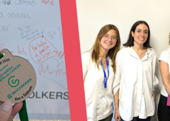
Ana Rita Barbosa de Matos is a postdoctoral researcher at i3S, Portugal who received an EACR Travel Fellowship to visit and work at VHIO, Spain between...
About Us
The Cancer Researcher is an online magazine for the cancer research community from the European Association for Cancer Research.
The EACR, a registered charity, is a global community for those working and studying in cancer research. Our mission is “The advancement of cancer research for the public benefit: from basic research to prevention, treatment and care.”

 Summary of the findings