The EACR’s ‘Highlights in Cancer Research’ is a regular summary of the most interesting and impactful recent papers in cancer research, curated by the Board of the European Association for Cancer Research (EACR).

The list below appears in no particular order, and the summary information has been provided by the authors unless otherwise indicated.

Explore the links or use the ‘Next’ and ‘Previous’ buttons to navigate

  1. Blocking Genomic Instability Prevents Acquired Resistance to MAPK Inhibitor Therapy in Melanoma

2. Spatial epitope barcoding reveals clonal tumor patch behaviors

3. Non-viral precision T cell receptor replacement for personalized cell therapy

4. The ectonucleotidase CD39 identifies tumor-reactive CD8+ T cells predictive of immune checkpoint blockade efficacy in human lung cancer

5. Genetic and pharmacological modulation of DNA mismatch repair heterogeneous tumors promotes immune surveillance

6. Loss of NECTIN1 triggers melanoma dissemination upon local IGF1 depletion

7. The NALCN channel regulates metastasis and nonmalignant cell dissemination

8. Bone Metastasis Initiation Is Coupled with Bone Remodeling through Osteogenic Differentiation of NG2+ Cells

9. Dynamics of age- versus therapy-related clonal hematopoiesis in long-term survivors of pediatric cancer

10. Genome-wide analysis of aberrant position and sequence of plasma DNA fragment ends in patients with cancer


1Blocking Genomic Instability Prevents Acquired Resistance to MAPK Inhibitor Therapy in Melanoma

Dharanipragada, P., Zhang, X. et al. Cancer Discovery. 13 (4): 880–909. (2023).
doi: 10.1158/2159-8290.CD-22-0787.

Summary of the findings

Acquired therapy resistance is mechanistically heterogeneous and, once clinically evident, liable to enhanced metastatic potential. Thus, research into specific vulnerabilities of acquired resistance must be complemented by understanding the origins of resistance (preexisting or de novo), which may rationalize preventive strategies. In this Cancer Discovery paper, the authors focused on the cancer where MAPK inhibitor (MAPKi) therapy was first developed, metastatic cutaneous melanoma, analyzed BRAFV600MUT melanoma, and extended their analysis to NRASMUT melanoma (where MAPKi-based therapy awaits development). Analysis of whole-genome sequences (WGS) delineated pervasive genomic instability, including chromothripsis. Comparative WGS analysis of patient-matched sensitive and acquired-resistant tumors revealed a specific pathway of genomic instability, gene amplification by extrachromosomal DNAs (ecDNA) and intrachromosomal complex genomic rearrangements (CGR). While ecDNAs and CGRs are present commonly in non-treated/sensitive tumors, they are distinct in acquired-resistant tumors (in their cargo genes and regulatory sequences, mutational signatures) and amplify bona fide resistance-driver genes (e.g., BRAF, NRAS, CRAF). Chromothriptic and ecDNA/CGR genomic spans overlapped non-randomly, suggesting chrommothripsis as a cause of ecDNAs/CGRs. Analysis of breakpoint-junctional sequences inferred non-homologous end-joining (NHEH) as key to ecDNA/CGR biogenesis. Targeting DNA-PKCS, a key NHEJ enzyme, prevented acquired MAPKi resistance in BRAFV600MUT and NRASMUT melanoma by limiting MAPKi-elicited ecDNA–CGR expansion.
Treatment of melanoma with MAPKi leaves persisters that later give rise to acquired resistance. Persisters may resume slowly cell cycling, with mitotic errors (e.g., lagging chromosomes, not shown) giving rise to micronuclei that enclose mis-segregated chromosomes or chromosome fragments. Micronuclei membranes are prone to rupture, exposing content chromosomes to mutagenesis and genomic instability, including chromothripsis. Genomic DNAs that drop out during re-ligation of chromothriptic fragments can circularize. Amplification of cargo genes and regulatory sequences ensue as a result of non-Mendelian inheritance of ecDNAs. Upfront DNA-PKi co-treatment attenuates ecDNA and CGR generation and prevents clonal diversification optimal for efficient escape from MAPKi.

Future impact

The immediate translational concept consists of the combination of MEKi plus DNA-PKi to treat NRASMUT melanoma, as MEKi monotherapy did not achieve clinically meaningful responses in this patient population. The same rationale may also justify clinical development for BRAFMUT or KRASMUT cancers. The notion of ecDNAs as master epigenomic reprogrammers of acquired resistance requires intense scrutiny for therapeutic opportunities. This work should spur studies to stabilize the cancer genome with the intent to prevent acquired therapy resistance. Targeting chromothripsis–ecDNA–CGR biogenesis may involve alternative pharmacologic approaches, such as mitigating chromosome segregation errors and rupture of micronuclei membranes.
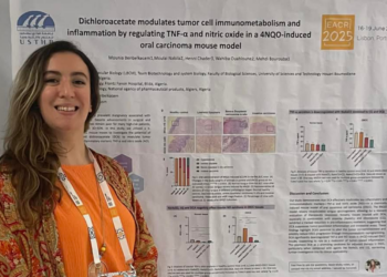
2Spatial epitope barcoding reveals clonal tumor patch behaviors

Rovira-Clavé, X., Drainas, A., P., Jinag, S., et al. Cancer Cell. 40 (11): 1423-1439. (2022).
doi: 10.1016/j.ccell.2022.09.014.

Summary of the findings

Intratumoral heterogeneity is associated with tumor progression and lack of response to treatment in patients with cancer. Despite advances in single-cell technologies, accurately tracking phenotypes and clonal evolution in situ within tumors remains a challenge. To address this issue, we have developed epitopes for imaging using combinatorial tagging (EpicTags). This tool was coupled with multiplexed ion beam imaging (MIBI) to create EpicMIBI, a technology that enables subcellular imaging of 40 proteins and in situ tracking of barcodes within tissue microenvironments. We used EpicMIBI to examine the spatial components of cell lineages and phenotypes in xenograft models of small-cell lung cancer; these tumors typically contain neuroendocrine (NE) and non-neuroendocrine cells (non-NE). We discovered that a small population of the non-neuroendocrine cells expanded to form large clonal patches, whereas neuroendocrine cancer cells did not. In addition, regions within the tumor had unique architectures; some contained large clonally diverse cell patches, and in other regions, the clonally diverse cell patches were smaller and disseminated. Lastly, epicMIBI can track the effects of genetic manipulations in subpopulations of cancer cells within a tumor. In a tumor model containing PTEN-deficient cancer cells, we observed a non-autonomous increase of clonal patch size in PTEN wild-type cancer cells.

Future impact

EpicMIBI allows tracking of phenotypes and clonal evolution in situ and enables investigation of both cell-intrinsic and cell-extrinsic processes involved in intratumoral heterogeneity. Combined with the ability to track hundreds of genetically modified cells, future work employing this technology will allow identification of causal relations between genotypes and phenotypes in the tumor microenvironment including the cancer-cell compartment and the stromal and immune cell compartments. This technique is a valuable tool for understanding tumor evolution and response to treatment and may ultimately lead to the development of more effective, personalized cancer therapies.

3Non-viral precision T cell receptor replacement for personalized cell therapy

Foy, S., P., Jacoby, K., Bota D., A. et al. Nature. 615: 687–696. (2023).
doi: 10.1038/s41586-022-05531-1.

Summary of the findings

In theory, every patient with cancer carriers their own cure. T cells in the body can specifically recognize and kill individual cancer cells through engagement of the T cell receptor (TCR). This first in-human phase I clinical trial demonstrated the clinical feasibility of truly patient-specific adoptive TCR T cell therapies for the treatment of patients with solid cancers. In order to do this, three novel core technologies had to be developed: 1) capture of tumour-specific T cells from the blood of each patient, followed by TCR isolation and validation for the selection of up to three unique patient tumour-specific TCRs 2) a non-viral precision genome engineering technology for TCR gene replacement of the original TCR with the tumour-specific TCR using CRISPR technology, and 3) a clinical grade manufacturing process for the generation of a multi-TCR T cell product of a defined composition. Sixteen patients were safely treated with their own personalized cell therapy in a dose-escalation trial; 5 had stable disease and 11 had disease progression. Post-treatment analysis demonstrated that T cells both persisted and trafficked to the tumour. Together, these technologies and Phase I clinical trial demonstrate the feasibility of patient-specific adoptive TCR-T cell therapies.
A personalized TCR T cell therapy starts and ends with the patient’s own cells. A tumour biopsy and blood sample are used to predict cancer specific mutations and generate a personalized peptide-HLA library to then capture an individual cancer-specific T cell from a patient’s own blood. T cells are then non-virally precision genome engineered to the replace the endogenous TCR with the cancer-specific TCR and Up-to-three unique neoTCR-products are manufactured and infused back into the patient. Image created with Biorender.

Future impact

This phase I clinical trial demonstrates for the first time, that a truly personalized TCR T cell therapy for the treatment of patients with solid cancer is feasible and safe. It provides the groundwork and a path forward that could open a new era in cancer therapy. With further development and optimization of the described novel technologies for HLA-inclusive T cell capture followed by TCR isolation and validation plus non-viral precision engineering for the manufacture of a personalized autologous cell product, personalized adoptive cell therapy could become a more widespread option for patients with cancer.

4The ectonucleotidase CD39 identifies tumor-reactive CD8+ T cells predictive of immune checkpoint blockade efficacy in human lung cancer

Chow A., Uddin, F., Z., Liu, M. et al. Immunity. 56 (1): 93-106. (2022).
doi: 10.1016/j.immuni.2022.12.001.

Summary of the findings

We performed single cell sequencing (scCITE/TCR/RNA) of T cells from lung cancer biospecimens in order to link protein CD39 expression with transcriptional profile and TCR sequence. Using TCR cloning, we demonstrated that CD39+ CD8+ T cells are enriched for tumor reactivity and that CD39 expression is a durable marker of TCR recognition of cognate peptide: MHC I on tumor cells. We observed that non-KRAS/HER2-altered lung adenocarcinoma (LUAD) tumors, which are typically not associated with tobacco use had lower CD39 expression on CD8+ T cells. In contrast, KRAS- and HER2-altered lung adenocarcinoma and squamous lung cancers had higher CD39 expression on CD8+ T cells. Surprisingly, small cell lung cancer tumors, respite their association with tobacco use and high tumor mutation burdens, had lower CD39 expression on CD8+ T cells. Finally, higher levels of CD39 expression on CD8+ T cells or higher transcriptional expression of a gene signature of CD39+ CD8+ T cells was associated with improved responses from immune checkpoint blockade therapy. Created with BioRender.com

The repertoire of tumor-infiltrating lymphocytes (TILs) can be vast, and many of these TILs are not endowed with tumor reactivity. While a number of reports have shown that tumor-reactive CD8+ TILs express CD39, few reports have demonstrated that conversely, CD39 can be leveraged to serve as a proxy of tumor-reactive CD8 T cells. Using single-cell CITE/RNA/TCRseq, we show that CD39+ CD8 T cells in human lung cancers demonstrate transcriptional and proteomic features of exhaustion, tumor reactivity, and clonal expansion. Moreover, T cell receptor (TCR) cloning revealed that CD39 expression enriched for tumor-reactive CD8+ T cell clones in human lung cancer biospecimens. We observed that expression of CD39 in human CD8+ T cells was driven by antigen-specific TCR stimulation that durably increased after antigen stimulation and was proportional to antigen density and TCR signal strength. Flow cytometry of 440 lung cancer specimens revealed that CD39 level on CD8+ T cells is only weakly correlated with tumoral features that currently guide lung cancer therapy, such as histology, driver mutation, PD-L1 and tumor mutation burden (TMB). Immune checkpoint blockade (ICB), but not cytotoxic chemotherapy, increased intratumoral CD39+ CD8+ T cells. Baseline CD39 levels on CD8+ T cells were associated with improved clinical outcomes, but not immune-related adverse events, from ICB therapy. Furthermore, a gene signature of CD39+ CD8+ T cells predicted benefit from ICB, but not chemotherapy, in a phase III clinical trial of non-small cell lung cancer. This comprehensive profiling of the clinical, pathological and molecular features highlights the utility of CD39 as a proxy of tumor-reactive CD8+ T cells in human lung cancer.

Future impact

The success of ICB is dependent on the presence of an adequate number of endogenous tumor-reactive CD8+ T cells. CD39 expression on CD8+ T cells represents a simple proxy for the size of this endogenous tumor-reactive repertoire that can prioritize ICB therapies for those patients most likely to benefit. Furthermore, CD39 expression on CD8+ T cells can be utilized to enrich for tumor-reactive TCR candidates that leveraged can be in adoptive T cell therapies.

5Genetic and pharmacological modulation of DNA mismatch repair heterogeneous tumors promotes immune surveillance

Amodio, V. et al. Cancer Cell. 41 (1): 196-209. (2023).
doi: 10.1016/j.ccell.2022.12.003.

Summary of the findings

Mismatch repair (MMR) activity is a key determinant of immune response in colorectal cancer. It is worth noting that MMR deficient (MMRd) tumors are categorized as “hot” tumors due to their ability to attract a significant immune infiltrate and exhibit highly responsiveness to immune checkpoint blockade inhibitors. Conversely, MMR proficient (MMRp) tumors are generally considered immunologically “cold” and have been shown to be resistant to immunotherapy. In a subset of colorectal cancer patients, both MMRp and MMRd subpopulations coexists in the same tumor, where potentially “cold” and “hot” components share the same microenvironment. However, the impact of MMR heterogeneity on tumor immune infiltration and immune surveillance remains incompletely understood. In this paper, Amodio and colleagues attempted to address this gap by modelling MMR heterogeneity in a syngeneic murine model using MMRp and MMRd tumor cells at varying proportion. They discovered that the injection of MMRp\MMRd heterogeneous tumors in syngeneic immunocompetent mice led to a reshaping of tumor immune environment that correlated with an increase in immune surveillance. Additionally, the authors demonstrated that treatments with 6 thioguanine, an antimetabolite agent capable of selecting for cells in which the MMR machinery is unfunctional, maximized the immune response against MMR heterogeneous murine tumors.

Future impact

The study by Amodio et al highlights the potential impact of MMR heterogeneity on immune response in colorectal cancer. The findings from the murine model suggest a need for further investigation into MMR heterogeneity in human samples and propose MMR modulation as a potential strategy to enhance immune surveillance in “cold” tumors.

6Loss of NECTIN1 triggers melanoma dissemination upon local IGF1 depletion

Ablain, J. et al. Nat Genet. 54: 1839–1852 . (2022).
doi: 10.1038/s41588-022-01191-z.

Summary of the findings

Our understanding of the mechanisms driving tumor dissemination is still incomplete, especially in non-epithelial tumors like melanoma. Only few genetic alterations have been shown to promote metastasis and non-genetic events have emerged as major contributors to cancer cell spreading. Here, we describe a cooperation between external stress and genetic abnormalities in the regulation of melanoma metastasis. We identified deletions of the adhesion gene NECTIN1 in approximately half of human melanomas and found that NECTIN1 levels were lower in metastases than in primary tumors. Through experiments in cell lines, zebrafish models and human melanoma samples, we discovered that NECTIN1 loss promotes melanoma dissemination specifically in response to a local drop in insulin-like growth factor 1 (IGF1) concentration. NECTIN1 is required for the formation of adherens junctions, cell-cell contacts typical of epithelial tissues. Surprisingly, IGF1 depletion elicited the robust formation of adherens junctions between NECTIN1-wildtype melanoma cells, suggesting that non-epithelial tumors can form epithelial structures under stress. In contrast, NECTIN1-deficient cells failed to establish these junctions and, instead, activated a cell-matrix adhesion program triggering their migration. These findings uncover a mechanism by which genetic alterations in cell-cell adhesion modulate the response of cancer cells to microenvironmental stress, thus controlling their metastatic behavior.

Future impact

Model for the role of NECTIN1 loss in melanoma dissemination. Image created with Biorender.

Recent evidence suggests a substantial overlap between the migratory state of cancer cells and the resistance to current anti-proliferative drugs. Our results indicate that the migratory state of cancer cells is regulated by a combination of different signals from their microenvironment, including chemical cues, such as growth factors, and physical conditions, such as the presence of neighboring cells to which they can adhere. A deeper understanding of the multiplicity of these signals and their associated pathways inside cancer cells may reveal new therapeutically actionable vulnerabilities of metastatic tumors.

7The NALCN channel regulates metastasis and nonmalignant cell dissemination

Rahrmann, E., P. et al. Nat Genet. 54: 1827–1838. (2022).
doi: 10.1038/s41588-022-01182-0.

Summary of the findings

Metastasis, the process by which cancer cells spread through the body, is the leading cause of cancer-related death. Despite decades of study, this process remains poorly understood and resistant to treatment. Metastasis has historically been assumed to be an abnormal process mediated by primary cancers. However, by using mouse models of gastric, intestinal and pancreatic cancer, we have divorced metastasis from upstream tumorigenesis and linked it to a natural physiologic process, which is central to the maintenance of normal tissues, uncovering a new paradigm for metastasis. We identified that genetic deletion or pharmacological blockade (GdCl3) of the Sodium Leak Channel Non-Selective Protein (NALCN) significantly increased epithelial cell dissemination into the blood from both normal and malignant tissues revealing this cascade as a cancer-independent phenomenon and a novel treatment target. NALCN loss- of-function in tumour-bearing mice significantly increased circulating tumour cell numbers in the peripheral blood and seeded metastases body-wide. In non-tumour-bearing animals, disseminated epithelial cells did not cause metastases but seeded in organs and apparently contributed to the normal structures of the organs such as renal tubules and glomeruli. These findings will allow refinement of the current model of metastasis and have unmasked a potential novel target for anti-metastatic therapies.
We have identified a single ion channel, NALCN, as a key regulator of cancer metastasis and non-malignant cell dissemination.

Future impact

Current chemotherapy and radiation fail to cure many cancer patients and tumours often relapse in a morphologically more aggressive and metastatic state, the primary cause of cancer-related deaths. Developing anti-metastatic therapies has proven difficult since potential therapeutic targets in primary tumours that drive metastases e.g., mutant oncoproteins, have proved hard to find. By divorcing the process of metastasis from oncogenic transformation we have uncovered a natural cell dissemination phenomenon that holds promise as a new target for anti- metastatic therapies.

8Bone Metastasis Initiation Is Coupled with Bone Remodeling through Osteogenic Differentiation of NG2+ Cells

Zhang, W. et al. Cancer Discov. 13 (2): 474–495 (2023).
doi: 10.1158/2159-8290.CD-22-0220.

Summary and graphical abstract by Dr. Alexandra Boitor, EACR Scientific Officer

Summary of the findings

A key experiment in this study made use of spontaneous metastasis mouse models where lung carcinoma cells were subcutaneously transplanted into mice. Once tumours reached 1 cm in diameter, tumours were resected, and the right femur bones were fractured by drilling or bending. Changes in tumour spread and distribution were noticed and proved to be dependent on NG2+ cells. This sparked interest in investigating the relationship between cell differentiation, proliferation and invasion in both homeostatic and pathogenic conditions. Vector images used for the development of this figure were taken from https://www.pngwing.com.

Various cancer types, including breast, lung, prostate, and colon cancers often metastasize to the bone and metastasis reoccurrence can be observed even years after tumour removal. The bone tissue and microenvironment are highly dynamic under both homeostatic and pathogenic conditions. In their study, Zhang et al. investigated the relationship between metastasis initiation and bone remodelling by making use of several pre-clinical cancer models including co-culture studies, 3D cell culture and mouse models for spontaneous bone metastasis. Results from their study identified NG2+ cells as modulators of osteogenesis and bone remodelling in homeostatic and fractured (but otherwise healthy) bone and facilitators of metastasis initiation and development in pathogenic conditions. Zhang et al propose that NG2+ cells and disseminated tumour cells directly interact in a unique way through heterotypic adherens junctions and this interaction promotes the proliferation and motility of cancer cells. At the core of this interaction, and therefore responsible for the pro-metastatic abilities gained by cancer cells was proposed to be N-cadherin expressed on NG2+ cells.  

Future impact

This study proposes a potential connection between bone remodelling and bone metastasis and may explain some circumstantial epidemiologic observations such as increased risk of metastasis in breast cancer survivors following bone fractures or metastatic spreading to the oral cavity and the jaws following dental implants in some patients diagnosed with breast or lung cancer.

Read more in Cancer Discovery

9Dynamics of age- versus therapy-related clonal hematopoiesis in long-term survivors of pediatric cancer

Hagiwara, K., Natarajan, S., Wang, Z., Zubair, H. et al. Cancer Discov. 13 (4): 844–857. (2023).
doi: 10.1158/2159-8290.CD-22-0956.

Summary of the findings

Somatic mutations in hematopoietic stem cells (HSCs) that confer growth advantage, known as clonal hematopoiesis (CH), are associated with the normal aging process. To investigate how cancer therapy may impact CH in pediatric cancer survivors who are at risk of accelerated physiological aging, the authors performed deep sequencing on 2,860 childhood cancer survivors (median follow-up time is 23.5 years) along with 324 age- and ethnicity-matched community controls. CH was detected in 15% of the survivors, a prevalence significantly higher than the 8.5% in community controls, and was associated with exposures to alkylating agents, radiation, and bleomycin. To differentiate age-related CH clones from therapy-related CH clones, the authors calculated the probability of a CH event occurring naturally at a specific age from the control cohort, which was then used to estimate the age-adjusted likelihood of developing CH for each survivor following a given cancer treatment. Therapy-related CH shows significant enrichment in TP53 as well as STAT3, a CH gene specific to Hodgkin lymphoma survivors. Single-cell profiling of peripheral blood samples from Hodgkin lymphoma survivors revealed STAT3 mutations to be predominantly present in T-cells and contributed by SBS25, a mutational signature known to be associated with procarbazine exposure. Serial-sample tracking reveals that larger clone size is a predictor for future expansion of age-related CH clones, while therapy-related CH remains stable decades post-treatment.

Future impact

Clonal hematopoiesis (CH) in childhood cancer survivors with long-term follow-up is attributable to both ageing and prior therapy exposure. CH mutations in STAT3 matched COSMIC signature 25 known to be associated with exposure to procarbazine, were detected in 10% of the Hodgkin lymphoma survivors and confer a growth advantage in T cells.

Despite the dramatic improvement in the 5-year survival rate in pediatric cancer over the last 50 years, survivors are still at risk of late effects associated with treatment. This first comprehensive CH analysis in long-term survivors of pediatric cancer identifies accelerated clonal hematopoiesis in pediatric cancer survivors, which is linked to both chemo- and radiotherapy. The contrasting dynamics of clonal expansion for age-related versus therapy-related CH suggest the need for longitudinal monitoring, which may involve genomic profiling at single cell level, to ascertain the long-term effects of therapy-induced CH in pediatric cancer survivors.


10Genome-wide analysis of aberrant position and sequence of plasma DNA fragment ends in patients with cancer

Budhraja, K.,K., McDonald, B., R., Stephens, M.,D. et al. SciTranslMed. 15 (678), 2023.
doi: 10.1126/scitranslmed.abm68.

Summary of the findings

Cell-free DNA in plasma is fragmented to a modal length of 167 bp, which corresponds to the length of DNA typically wrapped within a mono-nucleosome. Several groups have shown that the genomic locations of DNA fragments observed in plasma correspond to nucleosome positioning in lymphoid cells. More recently, the differences between nucleosome positioning across cell types are being leveraged to aid cancer detection. In this paper, Karan Budhraja, Bradon McDonald and colleagues hypothesized that compared to healthy individuals, plasma DNA from patients with cancer will have differences in end positions. To test this, they first identified a map of recurrently protected genomic regions using deep whole genome sequencing of plasma DNA from healthy individuals. Using this map, they measured the fraction of aberrant DNA fragment ends observed within protected regions in >2600 plasma samples from additional healthy controls as well as patients with cancer from across 11 subtypes. They found the fraction of aberrant DNA fragment ends was more common in patients with cancer, higher for mutated DNA fragments and higher for DNA fragments affected by copy number alterations. Combining this information with a sample-level analysis of nucleotide frequencies at fragment ends, they showed that a random forest machine model achieved 91% accuracy to distinguish between plasma samples from cancer patients and from healthy individuals.