Here are 3 of the most important things we learned at the EACR-OECI’s Molecular Pathology Approach to Cancer in Bergamo, Italy in March 2023.
Molecular pathology is revolutionising clinical practice in oncology and pathology, paving the way for precision medicine, and has become a rapidly growing area of research. This conference aimed to simulate the multidisciplinary teams that act in the real world to facilitate cancer research and to provide cancer care by addressing a diverse audience including pathologists and pathology residents, molecular pathologists, researchers in the field of molecular diagnostics and precision oncologists.
Various topics were covered in talks from this conference, including comprehensive cancer profiling in clinical diagnostics, upcoming applications of multiparametric immunoanalysis, novel approaches for developing and implementing biomarkers in oncology and pathology including liquid biopsy, tumour heterogeneity (spatial and longitudinal markers) and perspectives of artificial intelligence (AI) in pathology.
“Tumour evolution is not exclusively genomic” (Lukas Bubendorf)
Cancer genomics drove significant advances both in cancer care and research
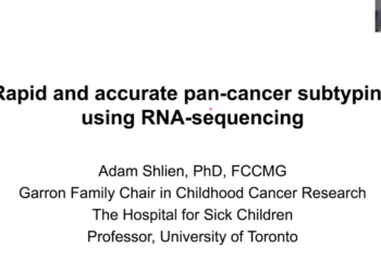
![]() It is well known by now that cancer develops as a result of accumulated genomic and epigenomic errors, and the development of next-generation sequencers allowed the cancer research community to reveal a large number of alterations in the genome of cancer cells. Nowadays targeted gene panels, whole exome and whole genome sequencing analysis of tumour samples inform tumour diagnosis, prognosis and treatment decisions, and also aid biomarker discovery and development of new therapies [1]. As comprehensive genomic analysis becomes more affordable, more and more clinical centres nowadays are looking into implementing tumour genotyping in patient care. To aid with this process during the conference, Dr Eli Pikarsky gave an interesting talk on “Establishing an In House Genomic Testing Facility”.
It is well known by now that cancer develops as a result of accumulated genomic and epigenomic errors, and the development of next-generation sequencers allowed the cancer research community to reveal a large number of alterations in the genome of cancer cells. Nowadays targeted gene panels, whole exome and whole genome sequencing analysis of tumour samples inform tumour diagnosis, prognosis and treatment decisions, and also aid biomarker discovery and development of new therapies [1]. As comprehensive genomic analysis becomes more affordable, more and more clinical centres nowadays are looking into implementing tumour genotyping in patient care. To aid with this process during the conference, Dr Eli Pikarsky gave an interesting talk on “Establishing an In House Genomic Testing Facility”.

Whilst cancer genomics drove significant advances both in cancer care and research, the fact that precision oncology nowadays focuses mostly on genetic mutations rises a few concerns. Some aberrations identified in the genome of cancer cells occur infrequently and generally speaking there’s a low rate of actionable mutations identified, with even fewer mutations ending up being targeted by genotype-matched drugs due to a lack of approved suitable medicine. In addition, challenges with variant interpretation commonly arise. Moreover, sometimes there’s a question if identified mutations are “true drivers” of cancer progression or just recurrent passenger mutations as the intratumoral heterogeneity is not accounted for in such analysis. Whilst predictive biomarkers are rapidly emerging, there’s still a question regarding the clinical utility of many of them. A solution to these shortcomings could be expanding tumour molecular profiling beyond genomics and making use of multi-omics approaches and tissue imaging techniques to achieve a better understanding of tumour biology [1].
Multi-omics approaches could improve treatment prediction
The use of transcriptomics data from tumour samples promises to improve several aspects of precision medicine, as shown by Dr Tuvik Beker and Dr Olli Carpen in their presentations. Dr Tuvik Beker and collab. developed ENLIGHT, a computational platform capable of predicting patients’ responses to cancer therapy based on cancer gene expression profiles. The ENLIGHT platform promises to improve treatment prediction for a large range of malignant diseases, including rare cancers and also to improve clinical trial design, as the use of this platform could prevent enrolment of patients unlikely to respond to the tested drug [2]. Moreover, Dr Beker went on to present ENLIGHT-DeepPT, a framework designed to predict mRNA expression based on H&E slides and accurately predict treatment response based on the estimated expression values [3].
In order to characterize chemotherapy resistance processes in high-grade serous ovarian cancer, Dr Olli Carpen and collab performed transcriptomic profiles at a single-cell resolution on paired tissue samples before and after chemotherapy. They noticed that chemotherapy promotes a stress-associated cell state. Interestingly enough, it seems that some cells are in this stress-associated state even prior to chemotherapy, with the treatment promoting a sub-clonal enrichment of this state. Dr Carpen and collab noticed a stress response not only in the tumour but also in the stroma, highlighting the fact that the development of chemotherapy resistance integrates stromal signalling and subclonal evolution [4].
Data on tissue heterogeneity and tumour microenvironment complement classic cancer genomics

In his presentation, Dr Carpen highlighted the impact that tumour heterogeneity can have on treatment response. In their studies, Dr Carpen and collab show that tumours display areas underlying relapse and chemoresistance characterised by specific tissue morphology [5, 6].
Dr Lukas Bubendorf’s presentation focused on the pronounced morphological and genomic intratumoral heterogeneity of non-small cell lung cancer (NSCLC) reflective of clonal evolution. Dr Bubendorf and collab showed that chromosomal instability characterises earlier stages of NSCLC and doesn’t increase during tumour progression [7-9]. He also discussed how tracking clonal evolution can explain the origin of metastases (which often don’t derive from the primary tumour) and the development of resistance to targeted treatments. Advances over the past decade were highlighted, and findings from the TracerX study were mentioned [10, 11].
Cell-cell interactions are a determining factor for cancer recurrence and survival, and as such profiling of functional proteins involved in cancer cell-stroma interactions could predict patient outcomes. In her presentation, Dr Leeat Keren showed how spatial context could be provided to protein expression in the tumour microenvironment through multiplexed imaging. Dr Keren’s work focuses on interactions involving immunoregulatory proteins, especially immune checkpoint expression patterns, and their prognostic role [12].
“AI is not taking over, it is just changing things” (Mara Graziani)
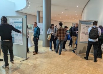
![]() Many of the talks presented at this conference focused on more technical aspects of research, especially data processing with several deep learning and AI-based technologies being discussed. For instance, apart from showing the correlation between PD-1 expression patterns and patient outcomes in triple-negative breast cancer [12], Dr Keren also discussed her computational analysis pipeline, the challenges she encountered and how deep learning could solve some of these challenges [13, 14]. Similarly, Dr Berek gave some insight into how the ENLIGHT platforms operate [2, 3].
Many of the talks presented at this conference focused on more technical aspects of research, especially data processing with several deep learning and AI-based technologies being discussed. For instance, apart from showing the correlation between PD-1 expression patterns and patient outcomes in triple-negative breast cancer [12], Dr Keren also discussed her computational analysis pipeline, the challenges she encountered and how deep learning could solve some of these challenges [13, 14]. Similarly, Dr Berek gave some insight into how the ENLIGHT platforms operate [2, 3].
Of note here was Dr Mara Graziani’s talk on “The human-AI horizon in microscopy imaging”. Dr Graziani began her talk with a comparison between the promises that AI brings and the Industrial Revolution and continued by highlighting the advantages that AI-based data analysis systems could bring to cancer care, but also the risks that implementation of insufficiently developed tools would bring. Dr Graziani stressed the need to ensure AI reliability and the importance of using large, high quality and diverse datasets alongside valuable expertise from domain experts that can provide examples of solid human-centric decision-making processes for the AI to learn from [15]. Further on, Dr Graziani presented her work on a piece of software that allows pathologists to inspect black-box classifiers in AI-analysed histopathology images [16].
The future lies in multidisciplinary approaches
![]() During the keynote lecture, Dr Judith Bovee presented an example of cancer where WHO has integrated molecular results with histology to improve the classification of disease stages, namely bone and soft tissue tumours. She also highlighted the importance of experienced multidisciplinary teams for the achievement of an accurate diagnosis. This allows for the classification to rely not only on histological and morphological traits but also on genetic data and presumed oncogenic mechanisms, revealing different levels of karyotype complexities and shared features of the different sarcomas [17].
During the keynote lecture, Dr Judith Bovee presented an example of cancer where WHO has integrated molecular results with histology to improve the classification of disease stages, namely bone and soft tissue tumours. She also highlighted the importance of experienced multidisciplinary teams for the achievement of an accurate diagnosis. This allows for the classification to rely not only on histological and morphological traits but also on genetic data and presumed oncogenic mechanisms, revealing different levels of karyotype complexities and shared features of the different sarcomas [17].
Whilst both the cancer research and cancer care communities agree on the benefits of precision cancer medicine, such approaches are difficult to implement in the clinic, especially in countries that rely on publicly funded healthcare systems. During this Molecular Pathology Conference, Dr Hege Russnes discussed the nationwide approach that Norway took to facilitate and promote the integration of cancer research and precision cancer medicine, particularly molecular pathology approaches in clinical practice, emphasizing the need of multidisciplinary teams comprised of oncologists, hematologists, pathologists and cancer researchers [18].
Dr Giorgio Stanta noted how far the Molecular Pathology field has come since 2003 when molecular pathology was first talked of at a clinical level and highlighted several European projects relevant to this research field. Dr Stanta also highlighted some of the shortcomings of the molecular pathology field, such as the lack of standardised ways to record and translate information between cancer centres and introduced OECI’s (Organisation of European Cancer Institutes) and UEMS’s (European Union of Medical Specialists) initiative to aid standardisation of procedures: a Master in molecular pathology.
The OECI-EACR Conference on Molecular Pathology Approach to Cancer also featured a panel discussion on “Unlocking the Potential of Molecular Pathology: Opportunities and Challenges Ahead”. [td_smart_list_end]

“The conference gave an updated view of current research across multiple countries in the field”
Participants praised the conference for the high scientific quality of the talks, friendly atmosphere and networking opportunities.
Overall this conference was an excellent opportunity for attendees to gain a comprehensive understanding of the scope, methods, future directions, and challenges in molecular pathology. Invited speakers and panellists discussed innovative approaches for empowering the next generation of molecular pathologists from both practical and educational viewpoints. The meeting fostered an environment that encouraged and facilitated participants to establish a network of interactions and to build bridges to foster cross-disciplinary studies.
Interested in EACR Conferences?
We organise a variety of excellent cancer research conferences, both in person and virtual, where the latest research topics and interaction for participants are the very highest priorities.
Make sure you add the dates of upcoming EACR Conferences to your diary now. Don’t forget we offer EACR member discounts on all of our registration fees!
Bibliography
1. Malone, E.R., et al., Molecular profiling for precision cancer therapies. Genome Medicine, 2020. 12(1): p. 8.
2. Dinstag, G., et al., Clinically oriented prediction of patient response to targeted and immunotherapies from the tumor transcriptome. Med, 2023. 4(1): p. 15-30.e8.
3. Danh-Tai, H., et al., Prediction of cancer treatment response from histopathology images through imputed transcriptomics. bioRxiv, 2022: p. 2022.06.07.495219.
4. Zhang, K., et al., Longitudinal single-cell RNA-seq analysis reveals stress-promoted chemoresistance in metastatic ovarian cancer. Science Advances. 8(8): p. eabm1831.
5. Laury, A.R., et al., Artificial intelligence-based image analysis can predict outcome in high-grade serous carcinoma via histology alone. Scientific Reports, 2021. 11(1): p. 19165.
6. Jamalzadeh, S., et al., QuantISH: RNA in situ hybridization image analysis framework for quantifying cell type-specific target RNA expression and variability. Laboratory Investigation, 2022. 102(7): p. 753-761.
7. Lorber, T., et al., Exploring the spatiotemporal genetic heterogeneity in metastatic lung adenocarcinoma using a nuclei flow-sorting approach. The Journal of Pathology, 2019. 247(2): p. 199-213.
8. Krause, A., et al., Deciphering the clonal relationship between glandular and squamous components in adenosquamous carcinoma of the lung using whole exome sequencing. Lung Cancer, 2020. 150: p. 132-138.
9. Krause, A., et al., Genomic evolutionary trajectory of metastatic squamous cell carcinoma of the lung. (2218-6751 (Print)).
10. Al Bakir, M., et al., The evolution of non-small cell lung cancer metastases in TRACERx. Nature, 2023. 616(7957): p. 534-542.
11. Frankell, A.M., et al., The evolution of lung cancer and impact of subclonal selection in TRACERx. Nature, 2023. 616(7957): p. 525-533.
12. Patwa, A., et al., Multiplexed imaging analysis of the tumor-immune microenvironment reveals predictors of outcome in triple-negative breast cancer. Communications Biology, 2021. 4(1): p. 852.
13. Greenwald, N.F., et al., Whole-cell segmentation of tissue images with human-level performance using large-scale data annotation and deep learning. Nature Biotechnology, 2022. 40(4): p. 555-565.
14. Elhanani, O., L. Keren, and M. Angelo, High-Dimensional Tissue Profiling by Multiplexed Ion Beam Imaging, in Single-Cell Protein Analysis: Methods and Protocols, A.T. Ooi, Editor. 2022, Springer US: New York, NY. p. 147-156.
15. Graziani, M., et al., A global taxonomy of interpretable AI: unifying the terminology for the technical and social sciences. Artificial Intelligence Review, 2023. 56(4): p. 3473-3504.
16. Graziani, M., et al. Sharpening Local Interpretable Model-Agnostic Explanations for Histopathology: Improved Understandability and Reliability. in Medical Image Computing and Computer Assisted Intervention – MICCAI 2021. 2021. Cham: Springer International Publishing.
17. Lam, S.W., T.M. Silva, and J.V.M.G. Bovée, New molecular entities of soft tissue and bone tumors. Current Opinion in Oncology, 2022. 34(4): p. 354-361.
18. Taskén, K., et al., A national precision cancer medicine implementation initiative for Norway. Nature Medicine, 2022. 28(5): p. 885-887.